

Translate this page into:
Oral cavity squamous cell carcinoma complicated by internal jugular vein thrombosis

* Corresponding author: Dr. Ashish Kumar Verma, Senior Resident, Department of Otorhinolaryngology- head and neck surgery, All India Institute of Medical Sciences, Bhopal, India. ashishverma4415@gmail.com
-
Received: ,
Accepted: ,
How to cite this article: Verma AK, Sahoo RK. Oral cavity squamous cell carcinoma complicated by internal jugular vein thrombosis. Future Health. 2024;2:166-9. doi: 10.25259/FH_48_2024
Abstract
Head and neck squamous cell carcinoma (SCC) encompasses various cancers from the upper aerodigestive tract. Initial presentation often includes regional nodal involvement (40% in stage IVA or B) and distant metastases (up to 10%). Facial edema is a frequent clinical issue with multiple causes, such as allergic reactions, trauma, venous thrombosis, infections, and both benign and malignant conditions. An extremely rare condition is tumor thrombosis of the internal jugular vein (IJV), typically reported in differentiated thyroid cancer cases. We present a diagnosed case of SCC of the left lower alveolus in a 54-year-old female who presented with diffuse facial swelling and stridor two years post-diagnosis. Further evaluation through clinical examination and imaging studies revealed partial thrombosis of the right IJV. The patient underwent an emergency tracheostomy and was treated with intravenous heparin, which effectively managed the local symptoms. This case report highlights the clinical features, diagnostic methods, treatment modalities, and favorable prognosis associated with the long-term effects of untreated head and neck cancers.
Keywords
Head and neck carcinoma
Facial edema
IJV thrombosis
Tumor thrombus
Tracheostomy

INTRODUCTION
Head and neck squamous cell carcinoma (HNSCC) describes various cancers that originate in the oral cavity, oropharynx, hypopharynx, and larynx. With over half a million cases reported each year, it is the fifth most prevalent cancer among men and the eighth most common cancer among women globally.1
Facial edema frequently occurs in patients with various cancers, including sarcoma, adenocarcinoma, and lymphoma. This cancer-related facial edema can be caused by factors such as superior vena cava syndrome, internal jugular vein thrombosis, and infections in the neck.2
Internal jugular vein (IJV) thrombosis is an extremely rare vascular condition often misdiagnosed or missed. It typically arises as a complication of intravenous drug abuse, extended use of central venous catheters, severe head or neck infections, hypercoagulable states, or trauma. While associated cancers are unusual and not extensively documented in the causes of IJV thrombosis, there have been a few reported cases of IJV tumor thrombus in patients with differentiated thyroid cancer.3 In this scenario, the situation becomes significantly more complex if SCC of the head and neck leads to the development of a tumor thrombus. We report a case of an elderly female diagnosed with SCC of the left lower alveolus, presenting with metastatic cervical lymphadenopathy and partial right IJV thrombosis.
CASE REPORT
The 54-year-old female initially presented with a non-healing ulcer in the left oral cavity two years ago, accompanied by pain. Examination revealed a proliferative growth in the left retromolar trigone area, which biopsy confirmed as moderately differentiated squamous cell carcinoma of the left lower alveolus. Fine needle cytology from a left level IB cervical lymph node indicated metastasis of squamous cell carcinoma. Imaging (Contrast enhanced computed tomography (CECT) neck) showed a heterogeneous lesion involving the left lower alveolus, extending into the ipsilateral retromolar trigone and causing cortical erosion of the mandible, along with an enlarged necrotic lymph node in the left level IB. Despite planning for surgery, the patient was lost to follow-up.
Two years after the initial diagnosis of carcinoma in the left lower alveolus, the patient presented with progressive facial swelling, painful diffuse neck swelling, dysphagia for solids, and dyspnea over 15 days. The swelling began insidiously in the neck and gradually extended cranially to involve the eyelids. The patient reported a recent onset of diminished vision in both eyes. Clinical examination revealed severe manifestations: the patient was in stridor. The face, lips, and eyelids showed non-pitting edema. A palpable 1×1 cm indurated, tender growth was noted in the oral cavity over the left retromolar trigone (RMT), along with tongue and lip edema. Neck examination demonstrated tense, hard, and tender diffuse swelling extending from the lower border of the mandible to the suprasternal notch, with restricted neck movements and obscured surface landmarks [Figures 1 and 2]
- Diffuse facial edema at the time of presentation.

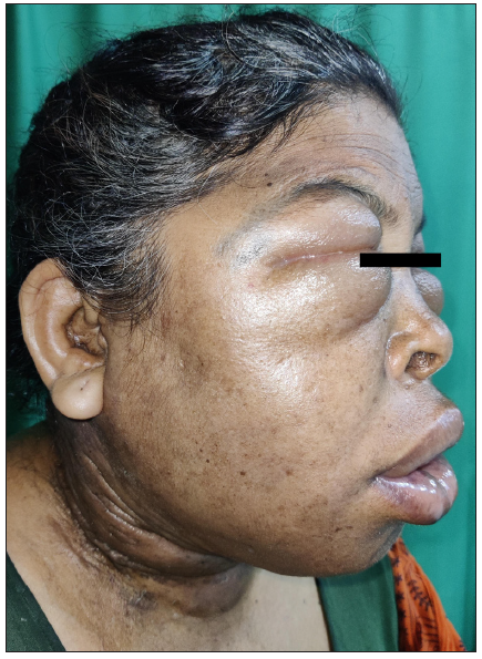
- Lateral view of the patient revealing edema involving eyelids, lip, face, and neck.
Fibre-optic laryngeal examination revealed narrowing of the laryngeal airway due to edematous swelling of the epiglottis, bilateral arytenoids, and pooling of secretions with evidence of aspiration; vocal cords were not visualized. USG color doppler revealed a partial thrombosis of approximately 5 cm in the right IJV, associated with necrotic lymph nodes and diffuse soft tissue thickening in the neck. Blood tests indicated metabolic alkalosis and electrolyte abnormalities (low sodium and potassium levels). Emergency management included performing a tracheostomy and initiating nasogastric tube feeding. The patient was subsequently admitted to the ICU, where treatment consisted of intravenous fluids, potassium supplementation, heparin, and dexamethasone. By the second postoperative day, there was a notable improvement in facial edema, and electrolyte balance was restored. Following clinical improvement [Figure 3], the patient was transferred to the ward, discharged on oral anticoagulants, and subsequently underwent radiotherapy as part of ongoing treatment.

- Post tracheostomy with resolved facial edema.
DISCUSSION
IJV thrombosis was initially documented by Long in 1912 in a patient with a peritonsillar abscess, and Goodman later suggested it could occur via lymphatics contaminated by infections in the oropharynx.4 Today, central venous catheterization is the leading cause, while other origins include Lemierre’s syndrome, malignancies, aneurysms, intravenous drug use, and idiopathic or iatrogenic factors.5
IJV thrombosis linked to malignancies can arise either from impaired venous flow caused by the tumor or metastatic deposits or from the hypercoagulable status frequently observed in cancer patients. Trousseau’s syndrome, characterized by recurrent or migratory thrombophlebitis in distant veins, is linked to various cancers, predominantly lung, pancreas, stomach, colon, and prostate. While most reported cases of tumor thrombus in the head and neck involve differentiated thyroid carcinoma, very few cases are associated with other head and neck malignancies such as buccal mucosa cancer, salivary carcinoma, or bronchogenic carcinoma. Wakasaki et al. were the pioneers in reporting cases of IJV thrombosis linked to SCC, highlighting its rarity outside of thyroid cancer.6 Recently, Tudor et al. reported a case of IJV thrombosis resulting from invasive pharyngeal cancer,7 while Cameron and colleagues described a case featuring necrotic cervical lymphadenopathy with IJV thrombosis in a patient with metastatic SCC of an unknown primary site.8
Clinical signs of IJV thrombosis encompass tender swelling in the neck, a palpable lump, and possibly pyrexia. Complications can involve pulmonary or septic embolism, septicemia, facial edema, and pseudotumor cerebri. Differential diagnoses to consider are cellulitis, infections in the neck spaces, and deep-neck abscesses.
Treatment typically involves anticoagulation to prevent clot propagation and embolization. Thrombolytic agents like streptokinase or urokinase may be used cautiously due to increased bleeding risk compared to heparin. Surgical options such as thrombectomy or venous ligation are rarely indicated.9
CONCLUSION
IJV thrombosis is an uncommon yet serious condition frequently linked to underlying malignancy. Early detection and swift treatment are essential to prevent potentially life-threatening complications.
Acknowledgements
I am deeply grateful for all the help and support I received throughout the completion of this case report. I sincerely thank Dr. Smriti for her invaluable support, advice, and insightful comments, which were instrumental in finalizing this report.
Ethical approval
Institutional Review Board approval is not required.
Declaration of patient consent
The authors certify that they have obtained all appropriate patient consent.
Financial support and sponsorship
Nil.
Conflicts of interest
There are no conflicts of interest.
Use of artificial intelligence (AI)-assisted technology for manuscript preparation
The authors confirm that there was no use of AI-assisted technology for assisting in the writing of the manuscript and no images were manipulated using AI.
REFERENCES
- Prolonged facial edema is an indicator of poor prognosis in patients with head and neck squamous cell carcinoma. Support Care Cancer. 2010;18:1313-9.
- [CrossRef] [PubMed] [Google Scholar]
- Deep neck infection as the main initial presentation of primary head and neck cancer. J Laryngol Otol. 2006;120:305-9.
- [CrossRef] [PubMed] [Google Scholar]
- Tumor thrombus of thyroid malignancies in veins: Importance of detection by ultrasonography. Thyroid. 2011;21:527-31.
- [CrossRef] [PubMed] [Google Scholar]
- Internal jugular vein thrombosis. Laryngoscope. 1985;95:1478-82.
- [CrossRef] [PubMed] [Google Scholar]
- Internal jugular venous thrombosis due to Trousseau’s syndrome as the presenting feature of metastatic prostate carcinoma: A case report. J Med Case Rep. 2016;10:104.
- [CrossRef] [PubMed] [PubMed Central] [Google Scholar]
- Massive internal jugular vein tumor thrombus derived from squamous cell carcinoma of the head and neck: Two case reports. Oral Maxillofac Surg. 2017;21:69-74.
- [CrossRef] [PubMed] [Google Scholar]
- Internal jugular vein thrombosis caused by invasive pharyngeal cancer: A case report and literature review. Braz J Otorhinolaryngol. 2024;90:101425.
- [CrossRef] [PubMed] [PubMed Central] [Google Scholar]
- Disseminated thrombosis of the internal jugular vein, superior ophthalmic vein and cavernous sinus as the primary manifestation of occult malignancy: A case report. Orbit. 2024;43:505-10.
- [CrossRef] [PubMed] [Google Scholar]
- Primary presentation of spontaneous jugular vein thrombosis to the otolaryngologist--in three different pathologies. J Laryngol Otol. 1998;112:888-90.
- [CrossRef] [PubMed] [Google Scholar]


